
Basics of Compound Microscope
Compound Microscope: Gateway to the Microscopic World
A compound microscope revolutionizes scientific observation by using multiple lenses to achieve magnifications up to 2000x. This powerful instrument enables researchers and students to explore bacteria, paramecium, blood cells, and countless other microorganisms with exceptional clarity.
Microscope View
An example of what you can observe under a compound microscope.
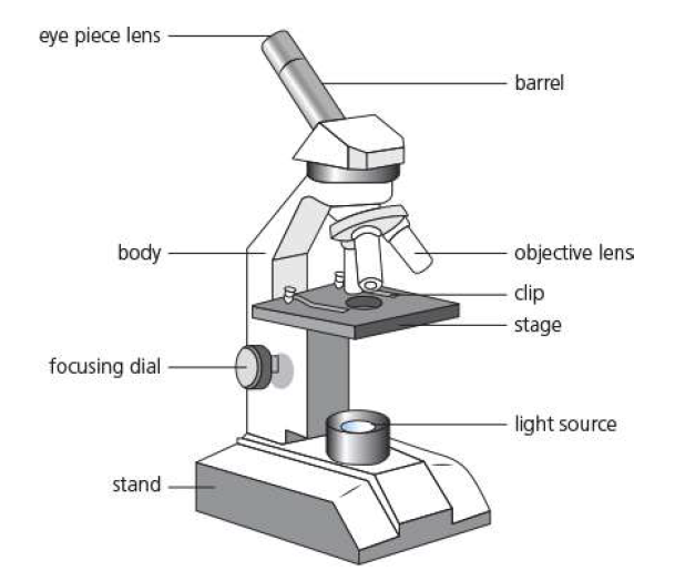
Microscope Parts Labeled and Compound Microscope Optical Parts
Understanding parts of a compound microscope and microscope labeled parts enables proper operation and maintenance. The label and parts of microscope systems include both mechanical and optical components essential for microscopy.
🔍 Ocular Lens (Eyepiece)
The lens you look through, typically providing 10x magnification. Modern compound microscopes feature high-quality ocular lenses for crystal-clear viewing. Essential component in parts of a microscope labeled diagrams.
🎯 Objective Lenses
Multiple lenses (4x, 10x, 40x, 100x) mounted on a rotating nosepiece. These compound microscope optical parts determine primary magnification for observing different organisms and represent key parts of a compound microscope.
🏗️ Stage and Microscope Glass Slides
The platform where specimens on microscope glass slides are placed. Features stage clips to secure slides and mechanical controls for precise positioning during microscope labeling exercises.
💡 Illumination System
LED or halogen light source with iris diaphragm for controlling light intensity and contrast. Critical component in microscope label diagrams for observing microorganisms effectively.
⚙️ Focus Controls
Coarse and fine adjustment knobs for achieving sharp focus. Essential parts of a microscope labeled in educational materials for observing bacteria, paramecium, and other microscopic organisms.
🔧 Iris Diaphragm Function
Controls light aperture and contrast in compound microscope parts. Proper diaphragm adjustment enhances visibility of transparent organisms like paramecium and euglena in microscope labeling studies.
Fascinating Organisms Under the Microscope
Microscope organisms reveal the incredible diversity of life invisible to the naked eye. From bacteria to complex protozoans, each organism displays unique characteristics and behaviors when observed through compound microscopes.
Bacteria Microscope Observations
Magnification needed: 400x-1000x
Bacteria appear as small, rod-shaped, spherical, or spiral organisms. Escherichia coli under microscope shows characteristic rod shapes and can be observed moving in liquid preparations. E coli bacteria under microscope display clear cellular boundaries.
Blood Cells Under Microscope
Magnification needed: 400x-1000x
Red blood cells under microscope appear as biconcave discs without nuclei, while white blood cells show various shapes and internal structures. Blood cells under microscope reveal different types including platelets as small fragments.
Plant Cell Under Microscope
Magnification needed: 100x-400x
Plant cells display rigid cell walls, chloroplasts, and large vacuoles. Onion cell under microscope shows clear cell boundaries and nuclei without chloroplasts, making them ideal for basic cell structure studies.
Cheek Cell Under Microscope
Magnification needed: 400x-1000x
Human cheek cells appear as large, flat, irregular shapes with visible nuclei. These animal cells lack cell walls and show flexible cell membranes, making them perfect for comparing with plant cells.
Sperm Cell Under Microscope
Magnification needed: 400x-1000x
Sperm cells under a microscope reveal distinct head, midpiece, and tail structures. Fresh samples show active motility, while stained preparations clearly display cellular components. Sperm cells under microscope demonstrate specialized cell morphology.
Spirogyra Under Microscope
Magnification needed: 100x-400x
Spirogyra displays characteristic spiral chloroplasts within filamentous cells. This green algae shows clear cell walls, nuclei, and the distinctive helical arrangement of photosynthetic organelles.
Yeast Microscope Observation
Magnification needed: 400x-1000x
Yeast cells appear as small, oval-shaped organisms that reproduce by budding. Active yeast cultures show parent cells with smaller buds attached, demonstrating asexual reproduction in real-time.
Pollen Under Microscope
Magnification needed: 100x-400x
Pollen grains display intricate surface patterns and shapes unique to each plant species. These microscopic structures show detailed textures and apertures essential for plant reproduction and identification.
Bone Under Microscope
Magnification needed: 100x-400x
Bone tissue reveals Haversian systems with central canals, concentric lamellae, and osteocytes in lacunae. Cross-sections show the organized structure that provides strength and support.
Specialized Microscopic Observations
🦠 Germs Under Microscope
Pathogenic microorganisms including bacteria, viruses (electron microscopy), and fungi. Proper staining techniques reveal cellular structures and help identify disease-causing organisms. Germs under a microscope show diverse morphologies.
🏥 Kidney Stones Under Microscope
Kidney stones under microscope reveal crystalline structures varying by composition (calcium oxalate, uric acid, struvite). Microscopic analysis helps determine stone type and treatment approach for optimal patient care.
🦠 Virus Under Microscope
Viruses require electron microscopy for visualization due to their nanometer size. Transmission electron microscope reveals viral capsids, envelopes, and internal structures at ultra-high magnification.
🪱 Tapeworm Under Microscope
Tapeworm segments show characteristic hooks, suckers, and reproductive organs. Cross-sections reveal internal anatomy including digestive and reproductive systems of these parasitic organisms.
🌊 Diatoms Under Microscope
Diatoms under a microscope display intricate glass-like shells with geometric patterns. These microscopic algae show remarkable symmetry and serve as indicators of water quality and environmental conditions.
🧠 Nervous Tissue Under Microscope
Nervous tissue reveals neurons with dendrites, axons, and cell bodies. Glial cells provide support structures, while synapses show connection points between nerve cells in neural networks.
🦴 Elastic Cartilage Under Microscope
Elastic cartilage shows chondrocytes within lacunae surrounded by elastic fibers. Found in ear and epiglottis, this tissue demonstrates flexibility while maintaining structural support.
🌿 Grass Under Microscope
Grass blade cross-sections reveal vascular bundles, chloroplast-containing cells, and specialized structures. Stomata on leaf surfaces control gas exchange in these monocot plants.
🍄 Mould Under the Microscope
Fungal hyphae form branching networks with spores and reproductive structures. Different mold species show distinct morphologies, spore arrangements, and cellular characteristics under microscopic examination.
💀 Human Ashes Under Microscope
Cremated remains show crystalline calcium phosphate structures and bone fragments. Microscopic analysis reveals mineral compositions and can help distinguish human from animal remains in forensic applications.
💇 Dandruff Under Microscope
Dandruff flakes appear as irregular keratin scales with possible fungal elements. Microscopic examination helps differentiate between dry skin, seborrheic dermatitis, and other scalp conditions.
Microscopy Calculations and Measurements
Problem 1: Total Magnification Calculation
Question: If you use a 10x eyepiece with a 40x objective lens, what is the total magnification?
Solution: 10x × 40x = 400x total magnification
Problem 2: Field of View Calculation
Question: If the field of view at 100x is 2mm, what is the field of view at 400x?
Solution: (2mm × 100x) ÷ 400x = 0.5mm field of view
Problem 3: Cell Size Measurement
Question: A paramecium spans 1/4 of the field of view at 200x magnification. If the field of view is 1mm, what is the actual size of the paramecium?
Solution: (0.25 × 1mm) ÷ 200x = 0.00125mm = 1.25 micrometers
Problem 4: Resolution Limit
Question: What is the theoretical resolution limit of a compound light microscope with numerical aperture of 1.4?
Solution: 550nm ÷ (2 × 1.4) = 196nm resolution limit
Types of Microscopes and Their Applications
Compound Microscopes and Complex Microscope Systems
Compound microscopes use multiple lenses to achieve magnifications up to 2000x. These complex microscope systems feature compound microscope parts including objective lenses, eyepieces, and illumination systems. Perfect for observing bacteria, paramecium, blood cells, and tissue samples with exceptional clarity.
Phase Contrast Microscope and Dark Field Microscope
Phase contact microscope enhances contrast in transparent specimens without staining, while dark field microscope illuminates specimens against dark backgrounds. Darkfield microscope techniques reveal living cells, bacteria, and microorganisms invisible in brightfield microscope observations.
Electron Microscope Technology
Electron microscope invention revolutionized scientific research by achieving nanometer resolution. Transmission microscope and scanning electron microscopes provide ultra-high magnification. Electron microscope price varies from $100,000 to over $1 million depending on specifications and capabilities.
Inverted Microscope Applications
Inverted microscope designs place objectives below specimens, ideal for cell culture work and live cell imaging. These instruments excel at observing cells in culture dishes and performing micromanipulation procedures.
🔬 Olympus Microscope Systems
Professional-grade microscopes known for superior optics and durability. Olympus offers compound, stereo, and digital microscopes for research and education with advanced camera and microscope integration.
🔬 Keyence Microscope Technology
Advanced digital microscopes with 4K imaging and measurement capabilities. Keyence microscope systems provide automated focus, measurement tools, and high-resolution documentation features.
📱 USB Microscope and Mini Microscope
Digital microscopes connecting directly to computers for real-time imaging. Mini microscope designs offer portability while USB microscope models provide educational demonstrations and basic research applications.
🔍 Microscope for Kindergarteners
Child-friendly microscopes with simple controls and durable construction. These educational instruments introduce young students to microscopy with safe, easy-to-use designs and basic magnification capabilities.
📷 Camera and Microscope Integration
Digital cameras designed for microscope attachment enable high-resolution image capture and documentation. Modern camera and microscope systems provide real-time imaging, measurement, and analysis capabilities.
💻 Digi-Microscope Systems
Fully digital microscopy platforms combining optics with computer processing. Digi-microscope technology offers automated imaging, measurement, and analysis with user-friendly software interfaces.
Microscope Components and Functions
🔧 Stage Microscope Function
The stage microscope function involves supporting and positioning specimens for observation. Mechanical stages provide precise X-Y movement, while stage clips secure microscope glass slides during examination.
🏗️ Arm Microscope Function
The arm microscope function provides structural support connecting the base to the optical components. This curved or straight support enables proper alignment and stability for accurate observations.
⚖️ Base Function in Microscope
Base function in microscope systems provides stability and houses electrical components. The heavy base prevents tipping while containing power supplies, controls, and illumination systems for optimal performance.
🔍 Microscope Lens Technology
Microscope lens systems include objectives and eyepieces with specific magnifications and numerical apertures. High-quality microscope lens designs minimize aberrations and maximize resolution for clear imaging.
🎯 Diaphragm Microscope Controls
Diaphragm microscope components control light intensity and contrast. Iris diaphragms adjust aperture size, while field diaphragms limit illumination area for optimal specimen visualization and contrast enhancement.
🔬 Contrast Microscope Techniques
Contrast microscope methods enhance specimen visibility through phase contrast, differential interference contrast, and polarization. These techniques reveal transparent structures without staining requirements.
Advanced Microscopy Techniques
Specimen Preparation for Optimal Viewing
Proper specimen preparation ensures clear observation of microorganisms. Wet mounts work best for living specimens like paramecium and bacteria, while fixed and stained preparations reveal internal structures.
Observing Sperm Through Microscope
Sperm cells require 400x-1000x magnification for detailed observation. Fresh samples show motility, while stained preparations reveal head, midpiece, and tail structures clearly.
Simple Squamous Epithelium Under Microscope
These thin, flat cells appear as irregular polygons with visible nuclei. Found lining blood vessels and air sacs, they demonstrate the relationship between structure and function.
Scientific References and Further Reading
NCBI – Microscopy Techniques in Cell Biology Nature Methods – Advanced Light Microscopy Nikon MicroscopyU – Educational Resources Olympus Microscopy Resource CenterFrequently Asked Questions About Microscopes
🚀 Explore More Scientific Subjects
Explore more biology topics like cell structure and biology experiments.